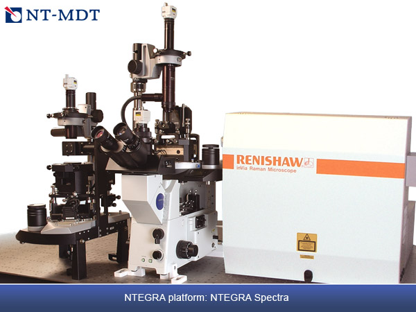
NTEGRA Spectra - AFM / CONFOCAL RAMAN & FLUORESCENCE / SNOM / TERS
Change happens at interfaces and today's most exciting changes in microscopy are happening where multiple technologies are interfaced together. NTEGRA Spectra is a prime example, uniting the full power of atomic force microscopy (AFM), confocal Raman and fluorescence microscopy and scanning near-field optical microscopy (SNOM) in one platform.
Simultaneous AFM and confocal Raman / Fluorescence imaging
NTEGRA Spectra supports most of the existing AFM modes (more than 30) providing comprehensive information about physical properties of the sample with nanometer scale resolution: local stiffness, elasticity, conductivity, capacitance, magnetization, surface potential and work function, friction, piezoresponse etc. Simultaneously with AFM, confocal Fluorescence and Raman measurements provide information about sample chemical composition, crystal structure and its orientation, presence of impurities and defects, macromolecular conformation, and so on. Measurements can be performed either through upright or inverted light excitation geometries. The sample can be in a controlled atmosphere or in a liquid environment, all under controlled temperature. Complete Raman /fluorescence spectrum is recorded in each point of 2D / 3D scan with further powerful software analysis. Due to the excellent microscopy performance of the NTEGRA Spectra, 3D spectral distribution can be studied with the spatial resolution reaching the theoretical limit.
Microscopy and spectroscopy at the molecular scale
Diffraction limited spatial resolution and weakness of Raman signal are the two major challenges in Raman microscopy. When using visible light, resolution of classical confocal microscopy does not go below 200 nm. The Raman signal is often only 1/millionth of the strength of a fluorescence signal. The new world of nanotechnology has disclosed a fascinating phenomenon: the electromagnetic field can be strongly enhanced near nanometer-scale metal asperities ("nano-antennas"). The resulting effects are called Surface Enhanced Raman Scattering (SERS) and, when done in conjunction with an SPM tip, one can get Tip-Enhanced Raman Scattering (TERS). By using a specially prepared sharp needle tip, NTEGRA Spectra can multiply the Raman signal strength by a few orders of magnitude from a precisely scanned, localized spot on the surface several nanometers in diameter. Even single molecules can be detected and recognized by their spectra. Lateral resolution of Raman (TERS) and fluorescence maps is no longer limited by light diffraction and can be less than 15 nm.
Specifications :
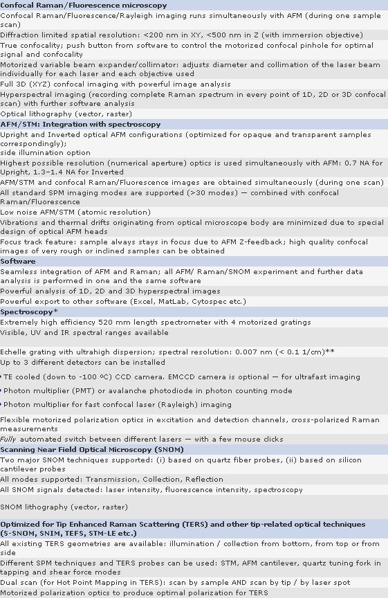
AFM-Raman measurements can run in air, in controlled atmosphere or in liquid - all with variable temperature
Some features listed are optional - not included into basic system configuration
* NT-MDT AFM can be integrated with Renishaw inVia or with NT-MDT spectrometer. Specifications are given for the latter. Renishaw specifications can be found at www.renishaw.com/AFM-Raman
** Exact value of spectral resolution highly depends on how "resolution" is defined
Applications :
- Graphene, carbon nanotubes and other carbon materials
- Semiconductor devices
- Nanotubes, nanowires, quantum dots and other nanoscale materials
- Polymers
- Optical device characterization: semiconductor lasers, optical fibers, waveguides, plasmonic devices
- Investigation of cellular tissue, DNA, viruses and other biological objects
- Chemical reaction control
|
|